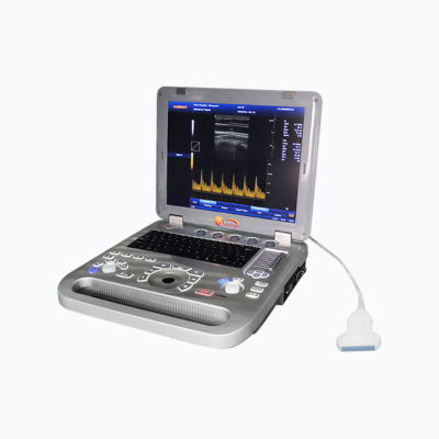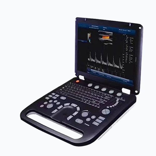
This advanced Color Doppler system features a large 15-inch LED display with real-time Doppler function, USB ports, VGA port, and two probe connectors. It includes a built-in battery offering up to 3 hours of continuous use without power. The system supports various imaging modes, including B, 2B, 4B, M, and Color Doppler options. It connects to printers of any brand and offers adjustable printed areas for pictures and reports. Additionally, it supports DICOM 3.0 protocol and provides multi-language support in English, Chinese, Spanish, Portuguese, Russian, Arabic, French, and more, making it a versatile diagnostic tool for medical applications.
Product Features
-
The system features a large 15-inch LED color display with real Doppler function, USB ports, and a VGA port.
-
It has a built-in battery, which can continuously operate for at least 3 hours when the power is off.
-
Main applications include Abdomen, Cardiac, Obstetrics, Gynecology, Urology, Andrology, Small Parts, Vascular, Pediatrics, Musculoskeletal, and more.
-
Imaging modes include B, 2B, 4B, M, B/M, B/C, B/D, B/C/D, B/CFM/D, CF+B model simultaneously, PDI Color, Dual Color, Simultaneous 2D/3D/4D Color Compound, PW, CW Duplex/Triplex, CFM, CDE, PD, Directional PD, CD, Anatomy M, and Color M mode.
-
The PC-based Color Doppler with Windows system can connect with any printer brand. The printed area is adjustable and can include pictures, reports, or a combination of both.
-
It supports the built-in DICOM 3.0 protocol.
-
It offers multi-language functionality in English, Chinese, Spanish, Portuguese, Russian, Arabic, French, Vietnamese, Indonesian, German, Persian, Thai, and more.
Product Specifications
| Weight | 5.5 kg |
|---|---|
| Dimensions | 37.5 × 36 × 7.5 cm |
| Doppler Mode | Pulsed Wave (PW) Doppler |
| Application | Cardiac, Musculoskeletal, Obstetrics, Small Parts, Urology, Vascular |
| Battery | Built-in Lithium Battery |
| DICOM Connectivity | DICOM 3.0 |
| Display Panel Technology | LED |
| Display Size(Diagonal) | 15-inch |
| Display Resolution | 1024×768 |
| Display Type | Color |
| Hard Disc | 120GB/200GB SSD (Optional), 64GB (SSD) |
| Imaging Mode | 2D, 3D, CF+B model simultaneously, DPDI, PDI, TDI, TSI |
| Instrument Classification | Class II |
| Material | Metal, Plastic |
| Place of Origin | Shanghai, China |
| Power Source | Electricity |
| Probe Connectors | 2 |
| PW Auto Trace Measurements | Yes |
| Quality Certification | CE (Conformité Européenne) |
| Safety Standard | None |
| Shelf Life | 2 Years |
| Software Package | General, OB/GYN, Small Parts, Urology |
| Warranty | 2 years |
- Connectivity Ports
- Display And Imaging
- Image Storage
- Transducers
- User Interface
- Performance Features
Connnectivity/Ports
| Transducer Ports |
2 |
|---|---|
| USB Ports |
2 |
| Ethernet Port |
2(100Mb/1000Mb) |
| External Display |
VGA, HDMI |
| Printer(Optional) |
USB Printer, Digital Laser Printer, Digital B/W Thermal Printer |
| Printing Area |
Image, Report, Image+Report |
| Footswitch |
USB |
Display & Imaging
| Display Type |
LED |
|---|---|
| Size |
15" (Diagonal) |
| Resolution |
1024x768 pixels |
| Contrast Ratio |
800:1 |
| Brightness |
230 cd/m² |
| Color Depth |
24-bit |
| Rotation Angle |
± 90° |
| Grey Levels |
256 |
| Cine Memory |
1200 frame (max) |
| Cine Review Speed |
1, 2, 4, 8 |
| Cine Review Loop |
Yes |
| Cine Capture Function |
Yes |
| Imaging Technologies |
Panoramic Imaging Technology, All-Digital Signal Processing Technology, Multi-Beam Formation Technology, Speckle Reduction Technology, Tissue Harmonic Imaging Technology, Dynamic Tissue Optimization Technology, Duplex & Triplex Synchronous Display, Directional Power Doppler, Imaging Parameters Preset, Tissue Special Image, PW Auto Trace, Update in Line, CF + B Mode on One Screen, Complex Model Imaging, IMT Auto Measurements, Virtual Convex Array, Trapezoidal Imaging |
| Image Zoom |
Real-Time Image Zoom |
| Zoom Range |
100%~400% |
| Image Inversion |
Flip the image along the Up/Down or Left/Right axis. |
| Pan Control |
Adjust the image position or flip it along any axis (up/down, left/right). |
Image Storage
| Storage Format |
PNG, AVI, BMP, JPEG, DICOM |
|---|---|
| Export Video Format |
AVI |
| Export Image Format |
PNG, BMP, JPEG, DICOM |
| External Storage |
USB Flash Drive |
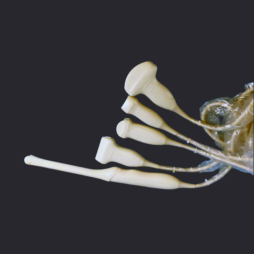
Transducers
| Probe Type | Frequency | Pitch | Radius | Elements |
|---|---|---|---|---|
| Convex Array Probe | Central 3.5 MHz (2.0 MHz to 5.0 MHz) | 0.516mm | 60 mm | 192 |
| Linear Array Probe | Central 7.5 MHz (5.0 MHz to 10.0 MHz) | 0.352mm | N/A | 192 |
| Intracavity Probe | Central 6.5 MHz (5.0 MHz to 8.0 MHz) | 0.216 mm | 10 mm | 192 |
| Microconvex Probe | Central 4.0 MHz (2.0MHZ to 10.0MHZ) | N/A | N/A | N/A |
User Interface
- User Permission Settings
- Intuitive Windows-based Operating Principles
- User-Centric Control Panel with Home Base Layout and Control Customization
- On/Off Task Light and Backlit Illumination of Control Panel
- Variable Brightness to Indicate Active State of Function Keys
- Easily Accessible, Full-Size QWERTY Keyboard for Text Entry, Function Keys, and System Programming
- Cine Playback, Multiple Arrows, Configurable Worksheets, Exam Review, Pictograms (Body Marks), System Setup Menu
- Online Attention Function, Guiding the User on How to Operate in the Next Step
Performance Features
| Digital Broadband |
12288 Channels |
|---|---|
| Beamformer |
Reprogrammable |
| Reprogrammable |
Adjustable (15 Steps) |
| Beamformer Frequency Range |
1-40 MHz |